Huntington’s disease (HD), a devastating neurodegenerative disorder, has long challenged scientists seeking to understand its underlying mechanisms. A breakthrough in research is emerging through advanced visualization techniques of Huntington’s protein aggregates, providing unprecedented insights into the disease’s progression. These protein clumps, formed by mutant huntingtin protein (mHTT), are central to HD pathology. Visualizing them is opening new doors for therapeutic development and early disease detection.
The Role of Huntington’s Protein Aggregates
Huntington’s disease is caused by a mutation in the huntingtin gene, leading to an abnormal expansion of CAG repeats. This results in the production of a mutated huntingtin protein that misfolds and aggregates, disrupting cellular functions. The formation of these Huntington’s protein aggregates is a hallmark of HD and contributes to neurotoxicity, neuronal death, and the hallmark symptoms of the disease, including motor dysfunction, cognitive decline, and psychiatric disturbances.
These aggregates are especially prevalent in the striatum and cortex, brain regions critical for motor and cognitive functions. Their presence has been linked to impaired cellular processes such as protein degradation, mitochondrial function, and synaptic transmission. Understanding their formation and impact is key to unlocking potential treatments for HD.
Visualizing the Invisible
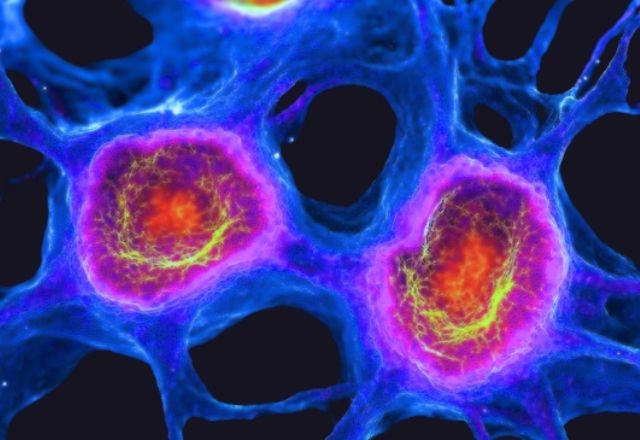
In the past, researchers faced challenges in studying Huntington’s protein aggregates due to their microscopic size and complex structure. However, advancements in imaging technologies are enabling scientists to visualize these aggregates at unprecedented resolutions. Techniques such as fluorescence microscopy, electron microscopy, and super-resolution imaging are providing detailed views of protein clumps in HD-affected cells.
- 1. Fluorescence Microscopy
Fluorescence microscopy uses fluorescent markers to highlight huntingtin aggregates in cells and tissues. These markers bind specifically to mutant huntingtin proteins, illuminating their structure and distribution. This method has helped identify the timeline of aggregate formation and their spatial relationship within affected neurons. - 2. Electron Microscopy
Electron microscopy offers high-resolution imaging to reveal the ultrastructural details of huntingtin aggregates. Researchers can observe their size, shape, and composition, aiding in the understanding of how these aggregates disrupt cellular architecture. - 3. Super-Resolution Imaging
Super-resolution imaging techniques, such as STORM and PALM, overcome the limitations of traditional optical microscopy, allowing visualization of nanoscale structures. Using these methods, scientists have uncovered intricate patterns of aggregate formation and interactions with other cellular components.
Key Discoveries from Aggregate Visualization
The ability to visualize Huntington’s protein aggregates is leading to significant discoveries:
Temporal Dynamics: Imaging studies have shown that aggregates form progressively, starting as small oligomers before evolving into larger inclusions.Cellular Localization: Aggregates are observed in both the cytoplasm and nucleus, suggesting they disrupt multiple cellular pathways.Interaction with Cellular Machinery: Advanced imaging reveals how aggregates interact with proteasomes, autophagosomes, and other cellular components, interfering with protein clearance and cellular homeostasis.
These findings are crucial for identifying potential intervention points where therapies could prevent aggregate formation or enhance their clearance.
Implications for Therapeutic Development
Visualization of Huntington’s protein aggregates is not just an academic exercise—it has direct implications for developing treatments.
Drug Screening
Advanced imaging enables researchers to screen potential drugs for their ability to reduce or prevent aggregate formation. Molecules that prevent huntingtin misfolding or enhance aggregate clearance can be tested in real time, accelerating drug discovery.
Gene Therapies
Gene-editing techniques, such as CRISPR-Cas9, are being developed to target the huntingtin gene. Visualization helps monitor the efficacy of these approaches by tracking aggregate reduction in treated cells.
Biomarker Development
Understanding the dynamics of protein aggregates aids in identifying biomarkers for early diagnosis. Imaging data can guide the development of non-invasive tests to detect aggregate presence in biofluids like cerebrospinal fluid.
Targeted Therapies
Antibody-based therapies and small molecules targeting aggregates are in development. Visualization techniques allow precise tracking of how these therapies interact with aggregates in cellular and animal models.
Challenges and Future Directions
While visualization offers immense promise, challenges remain. Aggregates are heterogeneous in size and composition, complicating their study. Imaging methods often require specialized equipment and expertise, limiting their accessibility. Additionally, while in vitro and animal models provide valuable insights, translating findings to human patients is a complex process.
Future research aims to address these challenges by:
Developing more accessible imaging platforms.Combining imaging with computational modeling to predict aggregate behavior.Expanding studies to include human-derived organoids and brain tissue.
Conclusion
The ability to visualize Huntington’s protein aggregates is revolutionizing our understanding of Huntington’s disease. By revealing the structure, formation, and impact of these aggregates, advanced imaging technologies are paving the way for novel diagnostics and targeted treatments. As research progresses, these tools may bring us closer to halting or even reversing the devastating effects of HD.
This intersection of cutting-edge technology and biomedical science offers hope to patients and families affected by Huntington’s disease, marking a significant step forward in the fight against neurodegenerative disorders.